Abdominal Anatomy Diagram / Abdominal Masses in the Neonatal Period | Pediatrics ... - Windham was previously a surgical oncologist in the sarcoma program of the h.
Abdominal Anatomy Diagram / Abdominal Masses in the Neonatal Period | Pediatrics ... - Windham was previously a surgical oncologist in the sarcoma program of the h.. Diagram of abdominal organs visceral organs advanced anatomy 2nd ed. The abdomen (colloquially called the belly, tummy, midriff or stomach) is the part of the body between the thorax (chest) and pelvis, in humans and in other vertebrates. Liver gallbladder pancreas labeled in male abdominal. Radiology basics of abdominal ct anatomy with annotated coronal images and scrollable axial images to help medical students and junior doctors learning anatomy. Lee moffitt cancer center & research institute in.
Diagram of abdominal organs introduction to the digestive system part 2 oesophagus and stomach 3d anatomy tutorial. Windham was previously a surgical oncologist in the sarcoma program of the h. But with the use of smart technology, you can learn faster and master abdomen anatomy in no sample decks: The stomach, the small intestine (jejunum and ileum), the large intestine (colon), the liver, the spleen, the gallbladder, the pancreas, the uterus, the fallopian. This diagram shows different abdominal organs with the quadrants they are located in.
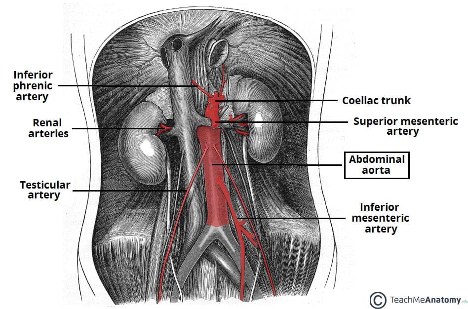
The Aorta - Branches - Aortic Arch - TeachMeAnatomy from teachmeanatomy.info Abdomen and digestive system anatomy: Kidneys are located retroperitoneally on the posterior abdominal wall on either side of vertebral column. Learn about its function, parts, abdominal conditions, and more. We focused especially on the diagrams of the abdominal digestive system (oesophagus is described on the modules about the thorax and oral cavity/pharynx. Regions abdominal abdomen region iliac anatomy anatomical pain left right inguinal area hypochondriac organs hypogastrium pelvic anterior upper cavity illustration. Loaded with beautifully illustrated diagrams clearly and concisely labeled for easy identification. Abdomen organs diagram, abdominal cavity organs diagram, abdominal organ anatomy quiz, internal abdominal organs diagram, upper abdominal diagram with ribs 12 photos of the abdominal diagram with ribs abdomen anatomy with ribs, abdominal anatomy with rib cage. Anatomy of the anterior abdominal wall and hernias, the gut and the peritoneal cavity, the retroperiteneum.
Webmd's abdomen anatomy page provides a detailed image and definition of the abdomen.
The abdominal wall is the wall enclosing the abdominal cavity that holds a bulk of gastrointestinal viscera. Introduction to sonographic abdominal anatomy. Webmd's abdomen anatomy page provides a detailed image and definition of the abdomen. Learn vocabulary, terms and more with flashcards, games and other study tools. Anatomy of the anterior abdominal wall and hernias, the gut and the peritoneal cavity, the retroperiteneum. Lee moffitt cancer center & research institute in. We focused especially on the diagrams of the abdominal digestive system (oesophagus is described on the modules about the thorax and oral cavity/pharynx. • in this module, we will explore basic abdominal anatomy identifiable with common imaging modalities. Not only can you answer questions asked directly about what is visible in an image, but you are able to apply the anatomy to sort of get your bearings in order to answer higher order questions. Abdomen and digestive system anatomy: A collection of anatomy notes covering the key anatomy concepts that medical students need to learn. Loaded with beautifully illustrated diagrams clearly and concisely labeled for easy identification. Lumbar spine pancreas ▫ anterior:
The bones of the abdomen are made up of the lumbar. Lumbar spine pancreas ▫ anterior: Abdominal anatomy, abdomen, gastrointestinal anatomy, gastrointestinal system. Diagram of abdominal organs visceral organs advanced anatomy 2nd ed. Diaphragm ▪ sac located beneath the liver ▫ inferior:
Abdomen Human anatomy Organ Human body, others free png ... from f0.pngfuel.com Kidneys are located retroperitoneally on the posterior abdominal wall on either side of vertebral column. Several key organs are packed closely with the stomach in the abdominal cavity, including the liver, whose smaller left. Lumbar spine pancreas ▫ anterior: Abdomen, abdomens, abdomen, abdominopelvis, abdominopelvic region, abdominopelvic regions, abdomen, abd, abdominal, abdominopelvic region, abdomen (volume), abdomen, nos. Regions abdominal abdomen region iliac anatomy anatomical pain left right inguinal area hypochondriac organs hypogastrium pelvic anterior upper cavity illustration. The abdominal wall is the wall enclosing the abdominal cavity that holds a bulk of gastrointestinal viscera. Sectional anatomy the sonographer must have a working knowledge of anatomical structures with particular the student in sonography needs to understand not only anterior to posterior anatomical structures, but also superior to inferior, medial to. Abdominal surface anatomy can be described when viewed from in front of the abdomen in 2 ways:
Anatomy of the anterior abdominal wall and hernias, the gut and the peritoneal cavity, the retroperiteneum.
Match each of the indicate the following body areas on the accompanying diagram by. Lumbar spine pancreas ▫ anterior: Abdomen and digestive system anatomy: Diagram of abdominal organs introduction to the digestive system part 2 oesophagus and stomach 3d anatomy tutorial. Lee moffitt cancer center & research institute in. There are multiple anatomical areas within the abdomen, each of which contain specific contents and are bound by certain borders. Muscular abdominal wall ▪ endocrine pancreas. Radiology basics of abdominal ct anatomy with annotated coronal images and scrollable axial images to help medical students and junior doctors learning anatomy. • in this module, we will explore basic abdominal anatomy identifiable with common imaging modalities. Describe the location, extent and gross features of kidney. Abdominal organ anatomy quadrants : Learn about its function, parts, abdominal conditions, and more. This mri abdomen axial cross sectional anatomy tool is absolutely free to use.
Several key organs are packed closely with the stomach in the abdominal cavity, including the liver, whose smaller left. Introduction to sonographic abdominal anatomy. Regions abdominal abdomen region iliac anatomy anatomical pain left right inguinal area hypochondriac organs hypogastrium pelvic anterior upper cavity illustration. Radiology basics of abdominal ct anatomy with annotated coronal images and scrollable axial images to help medical students and junior doctors learning anatomy. Human anatomy diagrams show internal organs, cells, systems, conditions, symptoms and sickness information and/or tips for healthy living.
Aorta - howMed from howmed.net Webmd's abdomen anatomy page provides a detailed image and definition of the abdomen. Sciency root words make anatomical parts harder to memorize. Several key organs are packed closely with the stomach in the abdominal cavity, including the liver, whose smaller left. Abdominal surface anatomy can be described when viewed from in front of the abdomen in 2 ways: Abdominal anatomy on computed tomography. Windham was previously a surgical oncologist in the sarcoma program of the h. Find this pin and more on human body anatomy diagram!!!! Sectional anatomy the sonographer must have a working knowledge of anatomical structures with particular the student in sonography needs to understand not only anterior to posterior anatomical structures, but also superior to inferior, medial to.
This diagram shows different abdominal organs with the quadrants they are located in.
This diagram depicts abdominal anatomy. The abdominal cavity is the part of the body that houses the stomach, liver, pancreas, kidneys, gallbladder, spleen, and the anatomy of the stomach area. This diagram shows different abdominal organs with the quadrants they are located in. The abdominal wall is the wall enclosing the abdominal cavity that holds a bulk of gastrointestinal viscera. Radiology basics of abdominal ct anatomy with annotated coronal images and scrollable axial images to help medical students and junior doctors learning anatomy. But with the use of smart technology, you can learn faster and master abdomen anatomy in no sample decks: Abdomen and digestive system anatomy: Learn vocabulary, terms and more with flashcards, games and other study tools. This mri abdomen axial cross sectional anatomy tool is absolutely free to use. The bones of the abdomen are made up of the lumbar. A collection of articles covering abdominal anatomy, including abdominal wall anatomy and abdominal cavity anatomy. Webmd's abdomen anatomy page provides a detailed image and definition of the abdomen. A good amount of area is covered by the abdominal wall.
Introduction to sonographic abdominal anatomy abdominal anatomy. The abdomen (colloquially called the belly, tummy, midriff or stomach) is the part of the body between the thorax (chest) and pelvis, in humans and in other vertebrates.